Difference between revisions of "Sylvain Faisan"
| Line 15: | Line 15: | ||
== Description of Activities == | == Description of Activities == | ||
| + | == Description des activités == | ||
| + | |||
| + | === Polarimetric Image Processing === | ||
| + | <table width=100%> | ||
| + | <tr> | ||
| + | <td> | ||
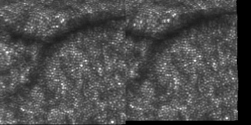
| + | We have proposed a new procedure to estimate polarization signatures for Stokes or Mueller images, while preserving sharp transitions. The procedure is based | ||
| + | on non-local means (NLM) filtering, which is an efficient denoising algorithm that outperforms | ||
| + | popular denoising methods regarding the preservation of sharp edges and fine texture details. | ||
| + | The noise is filtered while yielding physically admissible Stokes vectors (or Mueller matrices) at each pixel location. | ||
| + | The proposed joint filtering-estimation procedure is expressed as a constrained optimization problem. Interestingly, we show that it can | ||
| + | be equivalently seen as a two step method: a filtering stage based on the NLM approach followed by an estimation step ensuring physical admissibility. | ||
| + | Ellipticity of the Stokes vectors estimated with the proposed approach (left) and by using the pseudo-inverse (right) are presented. | ||
| + | * S. Faisan, C. Heinrich, G. Sfikas, J. Zallat, Estimation of Mueller matrices using non-local means filtering . Optics Express, pp. 4424--4438, Vol. 21, Num. 4, [http://dx.doi.org/10.1364/OE.21.004424 doi:10.1364/OE.21.004424], February 2013 | ||
| + | * S. Faisan, C. Heinrich, F. Rousseau, A. Lallement, J. Zallat, Joint filtering-estimation of Stokes vector images based on a non-local means approach . Journal of the Optical Society of America. A, Optics, Image Science, and Vision, pp. 2028--2037, Vol. 29, Num. 9, [http://dx.doi.org/10.1364/JOSAA.29.002028 doi:10.1364/JOSAA.29.002028], September 2012 | ||
| + | </td> | ||
| + | <td align=right> | ||
| + | [[File:polar.gif|frameless|thumb|upright=2.9]] | ||
| + | </td> | ||
| + | </table> | ||
| + | |||
| + | === Retinal Image Registration === | ||
| + | |||
| + | <table width=100%> | ||
| + | <tr> | ||
| + | <td> | ||
| + | We have proposed a new, robust and automated method for registering sequences of images acquired from scanning ophthalmoscopes. The method uses a multi-scale B-spline representation of the deformation field to map images to each other and an hierarchical optimization method. We applied the method to video sequences acquired from different parts of the retina. In all cases, the registration was successful, even in the presence of large distortions from microsaccades, and the resulting deformation fields describe the fixational motion of the eye. | ||
| + | * S. Faisan, D. Lara, C. Paterson, Scanning ophthalmoscope retinal image registration using one-dimensional deformation fields . Optics Express, pp. 4157--4169, Vol. 19, Num. 5, [http://dx.doi.org/10.1364/OE.19.004157 doi:10.1364/OE.19.004157], February 2011 | ||
| + | </td> | ||
| + | <td align=right> | ||
| + | [[File:animRecalage.gif|frameless|thumb|upright=2.9]] | ||
| + | </td> | ||
| + | </table> | ||
| + | |||
| + | === Comparing a subject to a statistical model === | ||
| + | <table width=100%> | ||
| + | <tr> | ||
| + | <td> | ||
| + | The goal of this project was to build a 3-D statistical model that represents the location of clinically relevant regions of the skull. The landmark distribution which is estimated from 3-D CT scans is modeled using a multivariate Gaussian Markov random field. The main contribution lies in a new way to characterize what constitutes an anomaly in a subject when it is compared to such a statistical model (which does not need to be a Gaussian Markov random field). Once global abnormality of the subject is detected, local anomalies are searched for by finding the smallest subset of landmarks whose well chosen displacement can render the subject normal according to the statistical model. | ||
| + | |||
| + | S. Faisan, A new paradigm to compare a subject to a statistical model. Application to the detection of skull abnormalities . Pattern Recognition Letters, pp. 1309--1315, Vol. 33, Num. 10, [http://dx.doi.org/10.1016/j.patrec.2012.03.009 doi:10.1016/j.patrec.2012.03.009], July 2012 | ||
| + | </td> | ||
| + | <td align=right> | ||
| + | [[File:model.jpeg|frameless|thumb|upright=2.9]] | ||
| + | </td> | ||
| + | </table> | ||
| + | |||
| + | === Warping a binary image... Simple, isn'it? === | ||
| + | <table width=100%> | ||
| + | <tr> | ||
| + | <td> | ||
| + | The estimation of one-to-one mappings is one of the most intensively studied topics in the research | ||
| + | field of non-rigid registration. Although the computation of such mappings can now be performed | ||
| + | accurately and efficiently, the solutions for using them in the context of binary image deformation is much | ||
| + | less satisfactory. In particular, warping a binary image with such transformations may alter its discrete | ||
| + | topological properties if common resampling strategies are considered. In order to deal with this issue, | ||
| + | this article proposes a method for warping such images according to continuous and bijective mappings, | ||
| + | while preserving their discrete topological properties (i.e. their homotopy type). Results obtained in the | ||
| + | context of atlas-based segmentation of complex anatomical structures highlight the advantages of the | ||
| + | proposed approach. At left, you can see the skull template that is warped in the proposed application (left, middle: front and profile view of the template. Right: template visualized with its topological skeleton). | ||
| + | |||
| + | * S. Faisan, N. Passat, V. Noblet, R. Chabrier, C. Meyer, Topology preserving warping of 3-D binary images according to continuous one-to-one mappings . IEEE Transactions on Image Processing, pp. 2135--2145, Vol. 20, Num. 8, [http://dx.doi.org/10.1109/TIP.2011.2158338 doi:10.1109/TIP.2011.2158338], 2011 | ||
| + | </td> | ||
| + | <td align=right> | ||
| + | [[File:ip2.jpg|frameless|thumb|upright=2.9]] | ||
| + | </td> | ||
| + | </table> | ||
| + | |||
| + | === Functional MRI - Brain Mapping === | ||
| + | |||
| + | <table width=100%> | ||
| + | <tr> | ||
| + | <td> | ||
| + | Activation detection at voxel v is formulated in terms of temporal alignment between sequences of hemodynamic response onsets detected in the fMRI signal at v and in the spatial neighborhood of v, and the input sequence of stimuli or stimulus onsets (see Fig. a). The multiple event sequence alignment problem is solved within the probabilistic framework of hidden Markov multiple event sequence models (HMMESMs). It consists in hypothesizing about a set of valid scenarios that could explain the N observed HRO event sequences. | ||
| + | A valid scenario (see Fig. b), relies on the combination of: | ||
| + | * an N + 1th event sequence, namely, a candidate sequence of task-induced hemodynamic activation onsets (HAOs) at the origin of the observations. | ||
| + | * HAO signatures, that is, associations of HRO events across channels, each association corresponding to the observable counterpart of a single HAO event. By definition, a signature is composed of one HRO event by observation channel, the event being observed (black line) or not (black point) (see Fig. b). | ||
| + | * Causality constraints between signatures given an HAO sequence. | ||
| + | If you are interested, you can find more information in the two following articles. In the second article, the neighborhood of v is not considered (N=1). | ||
| + | * S. Faisan, L. Thoraval, J.-P. Armspach, F. Heitz, Hidden Markov multiple event sequence models : a paradigm for the spatio-temporal analysis of fMRI data . Medical Image Analysis, pp. 1--20, Vol. 11, Num. 1, [http://dx.doi.org/10.1016/j.media.2006.09.003 doi:10.1016/j.media.2006.09.003], February 2007. | ||
| + | </td> | ||
| + | <td align=right> | ||
| + | [[File:problemStatement.jpg|frameless|thumb|upright=2.9]] | ||
| + | </td> | ||
| + | </table> | ||
== Publications == | == Publications == | ||
Revision as of 19:24, 20 March 2013
Assistant Professor
|
 |
Description of Activities
Description des activités
Polarimetric Image Processing
|
We have proposed a new procedure to estimate polarization signatures for Stokes or Mueller images, while preserving sharp transitions. The procedure is based on non-local means (NLM) filtering, which is an efficient denoising algorithm that outperforms popular denoising methods regarding the preservation of sharp edges and fine texture details. The noise is filtered while yielding physically admissible Stokes vectors (or Mueller matrices) at each pixel location. The proposed joint filtering-estimation procedure is expressed as a constrained optimization problem. Interestingly, we show that it can be equivalently seen as a two step method: a filtering stage based on the NLM approach followed by an estimation step ensuring physical admissibility. Ellipticity of the Stokes vectors estimated with the proposed approach (left) and by using the pseudo-inverse (right) are presented.
|
|

Retinal Image Registration
|
We have proposed a new, robust and automated method for registering sequences of images acquired from scanning ophthalmoscopes. The method uses a multi-scale B-spline representation of the deformation field to map images to each other and an hierarchical optimization method. We applied the method to video sequences acquired from different parts of the retina. In all cases, the registration was successful, even in the presence of large distortions from microsaccades, and the resulting deformation fields describe the fixational motion of the eye.
|
|
Comparing a subject to a statistical model
|
The goal of this project was to build a 3-D statistical model that represents the location of clinically relevant regions of the skull. The landmark distribution which is estimated from 3-D CT scans is modeled using a multivariate Gaussian Markov random field. The main contribution lies in a new way to characterize what constitutes an anomaly in a subject when it is compared to such a statistical model (which does not need to be a Gaussian Markov random field). Once global abnormality of the subject is detected, local anomalies are searched for by finding the smallest subset of landmarks whose well chosen displacement can render the subject normal according to the statistical model. S. Faisan, A new paradigm to compare a subject to a statistical model. Application to the detection of skull abnormalities . Pattern Recognition Letters, pp. 1309--1315, Vol. 33, Num. 10, doi:10.1016/j.patrec.2012.03.009, July 2012 |
|

Warping a binary image... Simple, isn'it?
|
The estimation of one-to-one mappings is one of the most intensively studied topics in the research field of non-rigid registration. Although the computation of such mappings can now be performed accurately and efficiently, the solutions for using them in the context of binary image deformation is much less satisfactory. In particular, warping a binary image with such transformations may alter its discrete topological properties if common resampling strategies are considered. In order to deal with this issue, this article proposes a method for warping such images according to continuous and bijective mappings, while preserving their discrete topological properties (i.e. their homotopy type). Results obtained in the context of atlas-based segmentation of complex anatomical structures highlight the advantages of the proposed approach. At left, you can see the skull template that is warped in the proposed application (left, middle: front and profile view of the template. Right: template visualized with its topological skeleton).
|
|

Functional MRI - Brain Mapping
|
Activation detection at voxel v is formulated in terms of temporal alignment between sequences of hemodynamic response onsets detected in the fMRI signal at v and in the spatial neighborhood of v, and the input sequence of stimuli or stimulus onsets (see Fig. a). The multiple event sequence alignment problem is solved within the probabilistic framework of hidden Markov multiple event sequence models (HMMESMs). It consists in hypothesizing about a set of valid scenarios that could explain the N observed HRO event sequences. A valid scenario (see Fig. b), relies on the combination of:
If you are interested, you can find more information in the two following articles. In the second article, the neighborhood of v is not considered (N=1).
|
|

Publications
<anyweb> http://newlsiit.u-strasbg.fr/papr/appli.php?author=faisan&title=&labo=tous&team=toutes&annee1=&annee2=&display=rap+&nationalRank=toutes&project=tous&hide=0&hide=0 </anyweb>