Sylvain Faisan
Maître de conférences
|
 |
Description des activités
Polarimetric Image Processing
|
We have proposed a new procedure to estimate polarization signatures for Stokes or Mueller images, while preserving sharp transitions. The procedure is based on non-local means (NLM) filtering, which is an efficient denoising algorithm that outperforms popular denoising methods regarding the preservation of sharp edges and fine texture details. The noise is filtered while yielding physically admissible Stokes vectors (or Mueller matrices) at each pixel location. The proposed joint filtering-estimation procedure is expressed as a constrained optimization problem. Interestingly, we show that it can be equivalently seen as a two step method: a filtering stage based on the NLM approach followed by an estimation step ensuring physical admissibility. Ellipticity of the Stokes vectors estimated with the proposed approach (left) and by using the pseudo-inverse (right) are presented.
|
|
Retinal Image Registration
|
We have proposed a new, robust and automated method for registering sequences of images acquired from scanning ophthalmoscopes. The method uses a multi-scale B-spline representation of the deformation field to map images to each other and an hierarchical optimization method. We applied the method to video sequences acquired from different parts of the retina. In all cases, the registration was successful, even in the presence of large distortions from microsaccades, and the resulting deformation fields describe the fixational motion of the eye.
|
|
Comparing a subject to a statistical model
|
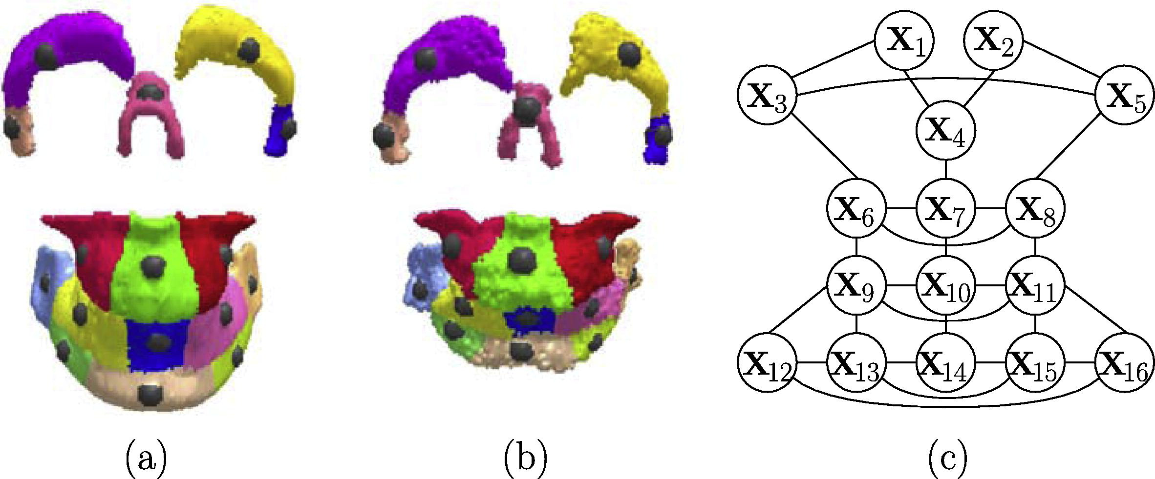
The goal of this project was to build a 3-D statistical model that represents the location of clinically relevant regions of the skull. The landmark distribution which is estimated from 3-D CT scans is modeled using a multivariate Gaussian Markov random field. The main contribution lies in a new way to characterize what constitutes an anomaly in a subject when it is compared to such a statistical model (which does not need to be a Gaussian Markov random field). Once global abnormality of the subject is detected, local anomalies are searched for by finding the smallest subset of landmarks whose well chosen displacement can render the subject normal according to the statistical model. The reference image and the regions of interest (each center of a sphere corresponds to the point (Fréchet mean) associated to a region) are presented in Fig. (a) whereas features extracted from a pathological subject are presented in (b). (c) is the dependency graph of the proposed Markov model. A node represents the position of a 3-D point. To make the relationship between the node and its associated point, the position of a node in the graph has been set to reflect the 3-D position of its associated point. |
|
functional MRI -- Brain Connectivity analysis
To do
Functional MRI -- Brain Mapping
|
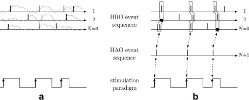
Activation detection at voxel v is formulated in terms of temporal alignment between sequences of hemodynamic response onsets detected in the fMRI signal at v and in the spatial neighborhood of v, and the input sequence of stimuli or stimulus onsets (see Fig. a). The multiple event sequence alignment problem is solved within the probabilistic framework of hidden Markov multiple event sequence models (HMMESMs). It consists in hypothesizing about a set of valid scenarios that could explain the N observed HRO event sequences. A valid scenario (see Fig. b), relies on the combination of:
If you are interested, you can find more information in the two following articles. In the second article, the neighborhood of v is not considered (N=1).
|
|
Publications
<anyweb> http://newlsiit.u-strasbg.fr/papr/appli.php?author=faisan&title=&labo=tous&team=toutes&annee1=&annee2=&display=rap+&nationalRank=toutes&project=tous&hide=0&hide=0 </anyweb>